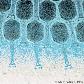
Long story short, phage display is a method used to find peptides that bind to proteins. You can also screen antibody fragments that bind to proteins. It's not a difficult research project. You buy a kit, buy or purify a protein and you go through the procedure. In the end you must verify that the peptide you found binds to your protein. Therein lies the wild west of phage display. You do not have a kit to tell you what is binding to what.
When a researcher starts a phage display project he/she has a loftier goal than fishing out binding peptides. Those peptides must also have a function. Most often that function is to inhibit the function of the protein they bind to. Let us look at one example of this work, keeping in mind that there are many others that make the same mistake. The mistake is made, it is published, and sometimes pursued as a drug candidate.
Several phage display projects have came up with peptides that are the same. The sequence SVSVGMKPSPRP has been found by many researchers. They further their findings by attaching a function that is done by the peptide. The problem is that this sequence is carried on a contaminating phage that comes from New England Biolabs PhD kits, lot 3. The phage can be found by anyone after several round of panning. Phage usually appear as blue dots in a lawn of bacteria. After several rounds you sometimes will see clear plaques. The clear plaques are the contamination. Early on NEB claimed that this came from sloppy lab techniques that resulted in contamination from phage floating around naturally in your lab. They have never admitted that any contamination exists in their libraries.
1 comment:
So helpful post. I found lots of information here. Keep it up!
Post a Comment